Your heart needs to be working correctly to maintain proper function without any issues that may result in future health problems. However, several issues like defects, blood clots, valve problems, and damage will make this proper functioning impossible. Thankfully, an echocardiogram Upper East Side can be beneficial in checking your heart structure and function and detect some of these issues before they progress further. There are three types of echocardiograms, and each works differently to give results. Therefore, your doctor will choose which one works well for you. Here is a discussion of the three main types of echocardiograms.
Transthoracic Echocardiogram
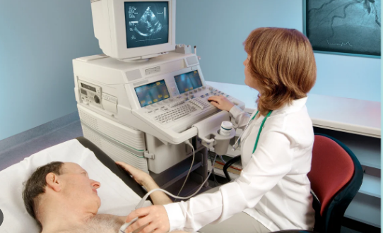
A transthoracic echocardiogram is the most commonly used type of echo, and your doctor performs it outside your body. You will not require any special preparations before your transthoracic echocardiogram. You will continue eating, drinking normally, and using your medications per their prescriptions. During a transthoracic echocardiogram, your sonographer places many electrodes on your chest and then attaches them to an electrocardiograph monitor. You may have to lie on your left side as your sonographer places a sound wave transducer on your chest with gel at its tip to help give clearer images. During the test, the transducer will pick up sounds, so you may hear swishing sounds that indicate your blood flow. Your sonographer may require you to hold your breath several times and move into different positions.
Transesophageal Echocardiogram
Unlike a transthoracic echocardiogram, a transesophageal echocardiogram takes images inside your chest and shows your heart and valves in greater detail. During this procedure, your sonographer will guide a transducer down your throat and esophagus. A transesophageal echocardiogram can give a detailed vision of your aorta and the back of your heart and evaluate your aortic and mitral valves. Also, it can check for blood clots and lung disorders. Additionally, this echo may be necessary if a transthoracic echocardiogram fails. Since a long tube will go down your throat during this procedure, your doctor will give you a solution to gargle so it numbs your throat. You will also require IV sedation to help you sleep throughout the procedure.
Exercise Stress Echocardiogram
A stress echocardiogram aims to show your heart’s working when it is taxed to tell how well it can withstand activity. First, your sonographer places electrodes on your chest and hook them to an electrocardiogram monitor to check your heart rhythm and rate during exercise. Next, your sonographer will measure your blood pressure, heart rate, and rhythm before starting your movement. Then, you will begin your exercise, probably on a treadmill, where you will increase intensity as you keep going. The exercise continues until you are exhausted. Meanwhile, your sonographer will ask about your symptoms while watching your heart on the monitor. However, if you cannot exercise on a treadmill for other reasons, your sonographer may give you medications to stress your heart.
An echocardiogram aims to reveal more details about your heart’s function and structure. Your doctor may recommend an echocardiogram to check your heart. If one is insufficient, your doctor will recommend another. After your echocardiogram procedure, you can ask your doctor to help you understand the meaning of your resulting images and if there are necessary steps. You can always ask your doctor about the procedure and if there are risks attached to them so you will be comfortable throughout the procedure. Generally, an echocardiogram is a safe procedure.